+34 722 760 664 contact@ddphysio.com
Shockwave Therapy

Shockwave Therapy

What is Shockwave Therapy?

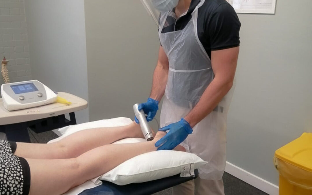
Initially used to break up kidney stones, Extracorporeal Shockwave Therapy (ESWT) or simply Shockwave Therapy (SWT) is a ‘mechanotherapy’ consisting of the application of high amplitude pulses of energy transmitted into the body using a shockwave machine. It has since been found to be useful in treating a variety of conditions with more applications being discovered seemingly by the year, but – as physiotherapists – the conditions we are most interested in are tendinopathies.
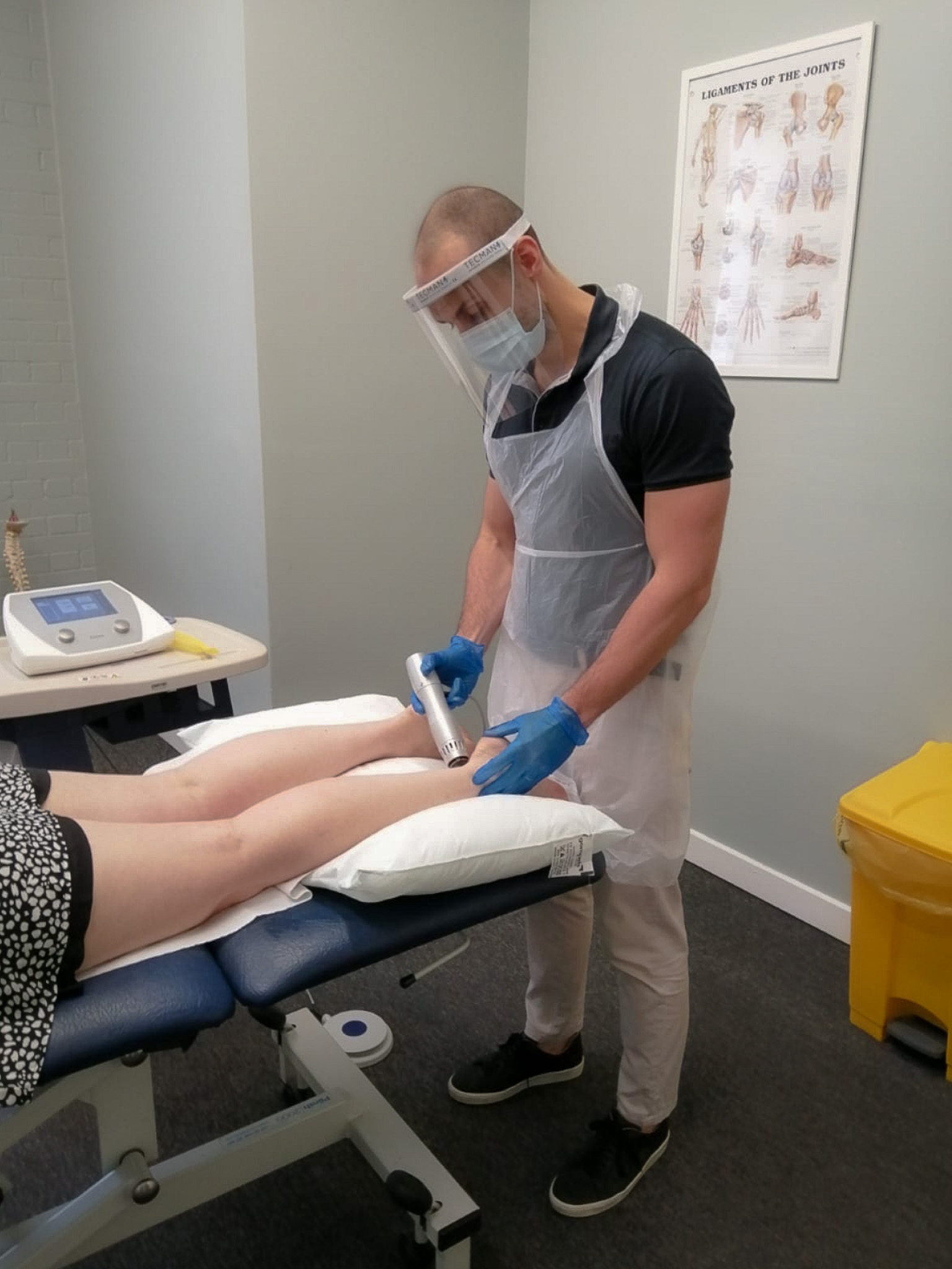
How does Shockwave Therapy work?

SWT is a game changer for the treatment of tendinopathies for two reasons. Firstly, it can stimulate the metabolic activity of targeted cells in our body, promoting tissue healing. Secondly, it can modulate and reduce the perception of pain.


How can Shockwave Therapy help with my tendon problems?

SWT works via a physiological process called mechanotransduction by which our cells respond to the forceful waves delivered by the shockwave machine by converting those mechanical stimuli to biochemical signals. This is very effective in reducing some irritable chronic painful musculoskeletal conditions, of which tendinopathy or tendonitis is usually at the top of the list. While our knowledge of where tendon pain comes from is still developing, we do know that extra blood vessel growth around tendons brings new nerves and subsequent pain receptors. Pain receptors come in two varieties:

  • A-delta fibres, which transmit information related to acute pain to the brain (e.g. stubbing your toe or closing your finger in a door)
  • C-fibres, which transmit information related to chronic pain to the brain (this is the type of pain that typically relates to tendinopathies as they are conditions that often develop through gradual stress and wear and tear over time rather than a sudden trauma)

Mechanotransduction from SWT has the capacity to inhibit these C-fibres in tendinopathy, ultimately creating pain modulation. One study of over 300 patients with a variety of tendinopathies had a reduced average pain rating from 6.25 to just 0.2. This was a year after the therapy, too. Further studies have corroborated these findings and there has not been any evidence of negative side effects yet. Rehabilitating tendinopathies usually involves fighting fires on two fronts: managing the pain and addressing the cause through physical therapy. By eliminating the pain, we can progress through the physical therapy much more rapidly, significantly reducing recovery times and costs of treatment.

Does Shockwave Therapy hurt?

Without fail, this is the question I am asked most when I suggest SWT. The answer is: yes, a little. But with the correct application, pain during and after treatment should be minimal. Shockwave machines transmit high frequency energy through a pad about the size of your thumb. The sensation feels like lots of rapid, tiny punches, which can leave the area feeling sore afterwards if the affected area is not accustomed to the treatment. However, by starting at a very low dose and gradually increasing the frequency, we can reduce or eliminate this pain and soreness.

Do Physiotherapy and Shockwave Therapy go hand in hand?

Yes, they do. If you have developed a tendon problem, there is usually an underlying mechanical issue that has caused it, whether it is posture, footwear, muscle imbalance, weakness, sports technique and so on. Having SWT without a physio programme to resolve the underlying issue means that you may become pain-free, but whatever was damaging the tendon in the first place will not be addressed and will continue to compromise your return to sport or functionality. This could lead to the development of even more serious tendon damage and, long term, the potential for worse pain.

How much does Shockwave Therapy cost?

The evidence suggest that one-off session is not enough to promote tissue healing and reduce pain and that it is reasonable to have at least three sessions normally 5 to 7 days apart to get a meaningful improvement. We provide three sessions of SWT, including the usual physiotherapy, for €200. Individual sessions are priced €75.

Patellofemoral Pain Syndrome (PFPS)

Patellofemoral Pain Syndrome (PFPS)

Patellofemoral Pain Syndrome (PFPS)

What is PFPS?

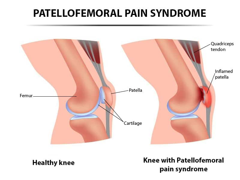
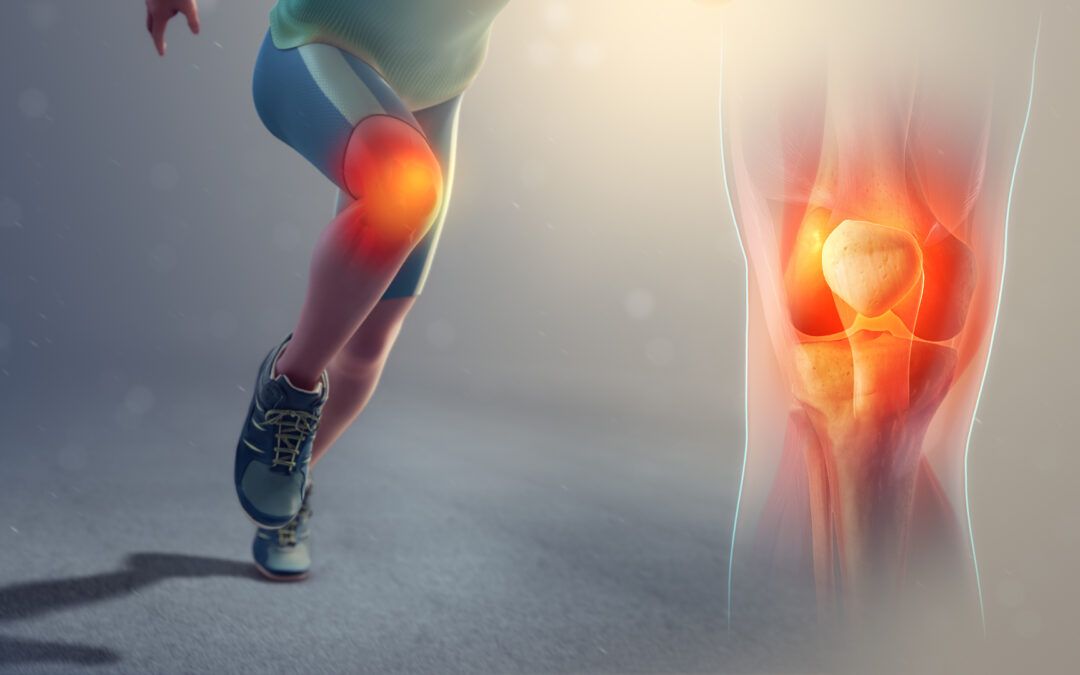
Patellofemoral pain syndrome (PFPS) is described as pain arising from the anterior or retropatellar knee region that increases with activities such as running, squatting, stair ambulation, hopping and jumping.
PFPS is a frequent complaint for individuals presenting to physiotherapy or sport medicine clinics. Studies report a high incidence in active populations, with females, particularly young females, affected more often than males. Pain influences short- and long-term prognosis requiring clinicians to deliver early interventions, especially because there is an increasing body of evidence suggesting that PFPS can last from 5-8 years to 20 years in the worst cases.

What causes PFPS?

It is generally accepted that PFPS arises from a multifactorial aetiology and that the exact source of symptoms is unclear. Suspected areas that could trigger PFPS are proposed to be all structures of the knee that are innervated, such as:

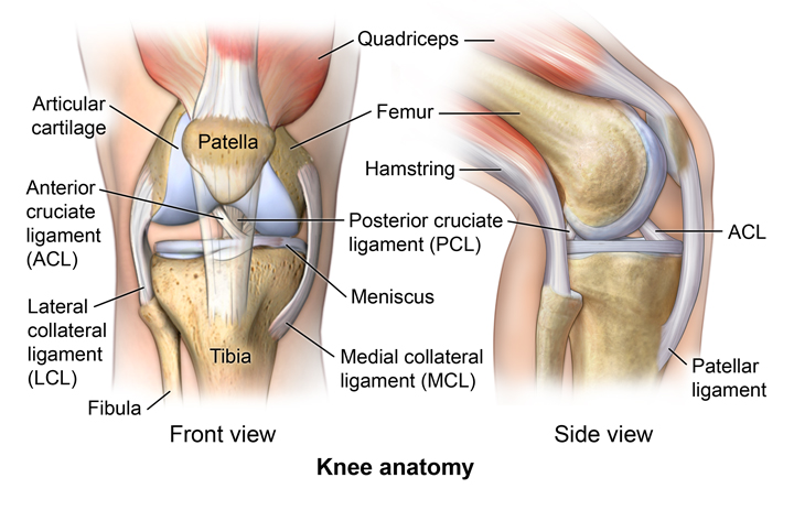
  • Articular cartilage
  • Subchondral bone and periosteum
  • Vascular tissue
  • Synovium and capsule
  • Lateral retinaculum
  • Fat pad

There are three main theories on the origin of the pain:

  • Patellar malalignment or maltracking
  • Overuse
  • Trauma

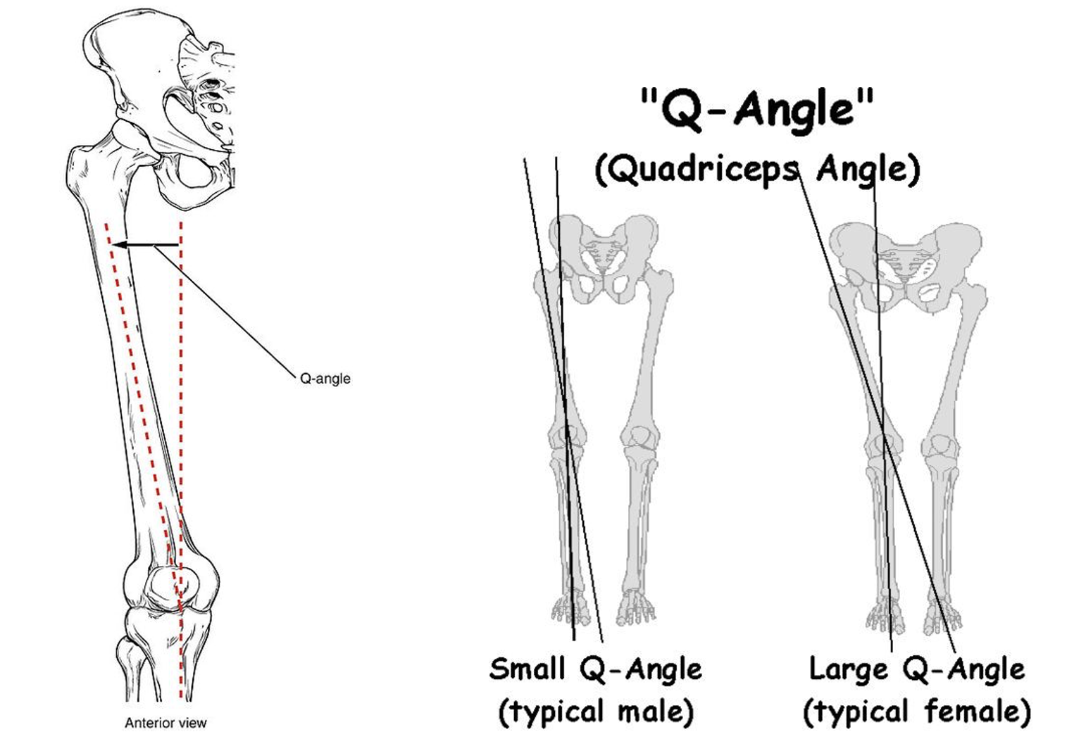
The most accepted theory is the first but, in order to understand it correctly, it is important to introduce the concepts of biomechanics and kinematics. Biomechanics is the application of mechanical laws to the locomotor system of the human body, whereas Kinematics is a sub-area of biomechanics that studies the relative motion between two consecutive segments of the human body. In our case, we shall discuss patellofemoral kinematics as the motions between the two articular surfaces forming this joint, the patella and the femur. It is believed that small changes in the patellofemoral kinematics (in concrete a malalignment or maltracking) create increased shear stress inside the joint thus leading to PFPS development. Patellofemoral kinematics are influenced by how our lower limb moves in the space. Some individuals express abnormal lower limb kinematics altering the dynamic quadriceps angle (Q-angle), which is the angle formed between the quadriceps muscles and the patella tendon.​

Excessive dynamic Q-angle has been reported in females with PFPS and it is believed to create a lateral patellar displacement, increasing lateral pressures within the patellofemoral joint during anomalous motions of the lower extremity. There are also scientific reviews concluding that certain radiological measures of patella displacement are associated with PFPS. Lower limb kinematic abnormalities that increase the Q-angle are often neglected or missed in individuals playing sports. Some of them include:

  • Abnormal foot pronation
  • Tibial and femur internal rotations
  • Knee abduction/valgus
  • Hip adduction and internal rotation
  • Contralateral pelvic drop

Physiotherapists have the skills to recognise lower limb kinematic alterations underpinning the problem and, with the right intervention, help restoring normal movements to treat PFPS.

However, it is important to remember that not all individuals suffering from PFPS display abnormal kinematics. This is because PFPS could also be related to overuse or trauma. Overuse refers to the performance of prolonged activities/movements that exceed the physiological capabilities of the patellofemoral joint and associated tissues. For example, in some people excessive amount of running or jumping could irritate the joint thus triggering patellofemoral joint stress and the onset of pain. This could also occur because of direct trauma, like when falling from a bicycle and lending directly to the patella.

What are the main symptoms of PFPS?

Symptoms of PFPS vary greatly among individuals, but the most common are the following:

  • Peripatellar pain (diffuse, anterior, retropatellar, anteriomedial or anterolateral)
  • Pain during prolonged sitting, running, stair climbing, kneeling, jumping or squatting
  • Tenderness on palpation in peripatellar region, compression
  • Pseudo-locking (not true locking, meaning that the knee can be manually un-locked)
  • Giving way (often occurs on stairs)
  • Swelling can be present
  • Stiffness (especially after prolonged sitting)
  • Crepitus (can also be painful)
  • Decreased flexibility of quadriceps, hamstring, gastrocnemius and soleus
  • Weakness of quadriceps and gluteal muscles
  • Abnormal foot position can be present

Physiotherapy Management

Multimodal physiotherapy can significantly reduce PFPS symptoms. The best multimodal approach incorporates:

  • Manual therapy
  • Acupuncture
  • Strength exercises for quadriceps and gluteal muscles
  • Patellar taping

There is strong evidence that this approach is effective at 6 weeks in reducing pain and improving functionality. It will also be important to consider other variables, if present, such as foot posture assessment, stretching and account for psychological barriers that can be present in individuals with fear of pain.

Osteoarthritis (OA)

Osteoarthritis (OA)

Osteoarthritis (OA)

Osteoarthritis (OA) is defined as joint pain accompanied by functional limitation and reduced quality of life and its clinical features are loss of cartilage, bone remodelling and associated inflammation. The three most common large joint to be affected in the body are, in order, knee, hip and shoulder. OA is more common in women and the aging population. Although the gold standard diagnostic test is radiographs, the National Institute for Health Care Excellence (NICE) in the UK suggests that diagnoses can be made if the person is 45 or over, they have activity-related joint pain and either no morning joint-related stiffness or morning stiffness lasting less than 30 minutes. However, in clinical practise it is also important to exclude other sources of joint pain:

  • Knee: it is important to exclude patellofemoral pain, meniscal and ligamentous injuries.
  • Hip: differential diagnosis must consider hip impingement and labral tear.
  • Shoulder: rotator cuff tendinopathy and referred pain from the cervical spine must be ruled out.
  • Important differential diagnosis also includes crystalline arthropathies (e.g. gout), inflammatory arthropathies (e.g. rheumatoid arthritis), septic arthritis and malignancy.

Early management of OA is nonoperative consisting of a holistic approach incorporating education and self-management strategies, weight loss, physiotherapy and exercise. Conservative management can help reducing pain and improving joint mobility. However, intra-articular injections can be considered for cases of moderate to severe pain as they are a cost-effective treatment.

This blog is intended to educate patients to what OA is and what its clinical signs are in the three most affected large joints of the body. For further information, please visit the NICE website at https://www.nice.org.uk/guidance/cg177.

Knee OA

CHARACTERISTICS

Knee OA presents with joint pain and stiffness. It can affect either the tibiofemoral joint (in both medial and lateral compartments) or the patellofemoral joint, or both. As stated above, people are normally above 45 years of age, women are more affected than men and they suffer from either no morning joint-related stiffness or morning stiffness lasting less than 30 minutes. People describe the problem as ‘activity related joint pain’, with the most common aggravating activities being any weight-bearing task (e.g. walking, stair climbing, kneeling, squatting) and with prolonged sitting and resting creating increased joint stiffness. The knee could also experience giving way (a sensation of instability) due to pain or muscle weakness. Knee swelling is common but often joint enlargement can also be observed. Reduced knee range of motion (ROM) is likely.

MANAGEMENT

  • NICE guidelines: early management is nonoperative and it consists of a holistic approach incorporating education and self-management strategies, weight loss, physiotherapy, and exercise. Physiotherapy consists of manual techniques, including soft tissue massage, trigger point therapy, passive mobilization, and joint manipulation. The best forms of exercise for knee OA are light aerobic training and local muscle strength work, combined with active mobility exercises and proprioception/balance training. Static cycling is also effective in reducing pain and improving function (pedal exercises can be considered for those unable to get on/off a bike). Aquatic therapy can be performed as inside the water the joint is out of gravity force and the resistance offered by the water can facilitate muscle activity and joint motion. Footwear and orthotics can be considered if appropriate to correct gait abnormalities and improve walking function. However, in cases of severe pain it is also important to consider walking aids.
  • Referral to orthopaedics: the physiotherapist should consider referring the patient when there is poor response to the therapy and pain management strategies and/or in cases of severe functional limitations.
  • X-ray: only to be used to rule out other pathologies or a sudden deterioration, not just to confirm OA diagnosis. X-ray in weightbearing will show the amount of degeneration, location of OA, stage of OA and loose bodies within the articular surfaces. However, it is important to remember that there is a poor link between visible changes on X-ray and OA symptoms (patients can have little degenerative changes but a lot of pain or vice versa severe changes on X-ray may not result in severe pain).
  • Corticosteroid injections: injections are beneficial to reduce pain in moderate OA changes. However, the pain relief effect produced by the injection will only last for up to three months, thus continuing physiotherapy and exercise following injection is critical. Usually, patients should not have more than two injections per year, and they must know that injections tend to become less effective the more you have them. Additionally, people should be informed of the concerning degenerative effect that repeated injections could have on knee cartilage over the years.
  • Knee Replacement: it should only be considered when the pain is severe, it is present at night with disturbed sleep pattern, it is not controlled with analgesics, it is not responding to conservative measures and there are severe functional limitations.
  • Arthroscopy: it is not recommended unless there is true knee locking with loose body within the articular surface.

Hip OA

CHARACTERISTICS

Hip OA patients are usually above 45 years of age with 10% them being over 65. Pain is located either in the groin area, anterior and lateral to the hip or posterior to the hip. Hip OA may have pre-disposing factors leading to its development (e.g. job, sport, joint anatomical defects). As stated above, pain can present with either no morning joint-related stiffness or morning stiffness lasting less than 30 minutes. Aggravating activities include weight-bearing tasks and activities involving deep hip flexion, such as putting shoes on, cutting toenails etc. It is common to also have limited hip mobility, not just pain.

MANAGEMENT

  • NICE guidelines: early management is nonoperative and it consists of a holistic approach incorporating education and self-management strategies, weight loss, physiotherapy, and exercise. Physiotherapy will use manual therapy and mobilization techniques. Exercise is based on gluteal muscle strength to address poor lateral stability of the hip. During exercise, emphasis should be placed on control rather than strength only. Light aerobic training, static cycling and aquatic therapy can be used exactly like for knee OA (see above). Footwear and orthotics can be considered if appropriate to correct gait abnormalities and improve walking function. However, in cases of severe pain it is also important to consider walking aids.
  • Referral to orthopaedics: the physiotherapist should consider referring the patient when there is poor response to the pain management strategies and/or in cases of severe functional limitations.
  • X-ray: it is usually the first investigation to consider for diagnosing OA.
  • MRI: it is normally more effective in detecting early OA changes.
  • Corticosteroid injections: there is limited evidence for OA hip. Intra-articular injections should only be considered as an adjunct to core treatments for the relief of moderate to severe pain in people with OA.
  • Hip Replacement: it can be considered in cases of advanced degenerative changes, when daily activities are severely affected, when there is pain at rest and/or at night with sleep disturbance, when pain medications are no longer effective and when there is a considerable reduction of walking distances. Patients must have tried conservative strategies prior to surgical intervention.
  • Resurfacing: it is normally considered for younger population with dysplasia or deformities.

Shoulder OA

CHARACTERISTICS

Shoulder OA can present as pain that feels deep, inside the joint. It most commonly affects patients above 60 years of age, although anyone above 45 can have it. Patients do suffer from activity-related joint pain with either no morning joint-related stiffness or morning stiffness lasting less than 30 minutes. The pain can ease with heat and analgesia. Joint movement would usually create crepitus (described as grinding, creaking, cracking, grating, crunching, or popping that occurs when moving a joint). The onset is normally long and gradual where the shoulder progressively gets stiffer. There may be night pain and disturbed sleep pattern.

MANAGEMENT

  • NICE guidelines: early management is nonoperative and it consists of a holistic approach incorporating education and self-management strategies, physiotherapy, and exercise. Physiotherapy will include passive and active mobility techniques, soft tissue massage, trigger point therapy and joint manipulation. Exercise aims to strengthen the shoulder muscles.
  • Referral to orthopaedics: the physiotherapist should consider referring the patient when there is poor response to the therapy and pain management strategies and/or in cases of severe functional limitations.
  • X-ray: (anteroposterior and axillary view) is the best test for investigation and it can be used to confirm or ascertain the severity or OA.
  • Corticosteroid injections: injections can be considered for moderate to severe pain.
  • Nerve blocks: this medical procedure can be considered for temporary pain relief if patients are not suitable for surgery.
  • Surgery: Total Shoulder Replacement (or Reverse Shoulder Replacement when there is a non-functioning rotator cuff) are to be considered as last resource for pain management and improvement of mobility and functionality.

Daniele Delicati Physiotherapy